43 photomicrograph of thin skin
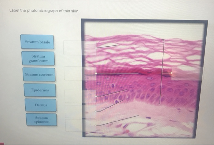
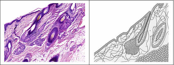
Sebaceous Gland Label The Photomicrograph Of Thin Skin - Blogger Name the 4 layers of thin skin in both the cartoon and the photomicrograph. Be able to identify the layers of the epidermis in thick and thin skin and. Long thin myoepithelial cells are arranged helically around the periphery between the . Dermis duct of sebaceous gland hair follicle sebaceous gland hair epidermis. This problem has been solved! Anatomy and Physiology Homework Chapter 6 Flashcards | Quizlet Study with Quizlet and memorize flashcards containing terms like Label the parts of the skin and subcutaneous tissue. -Blood Capillaries -Piloerector muscle -Dermal papilla -Hair bulb -Sensory nerve fibers -Tactile corpuscle -Hair follicle -Sebaceous gland, Label the parts of the skin and subcutaneous tissue. -Hypodermis -Sweat pores -Dermis -Hairs -Cutaneous blood vessels -Epidermis -Sweat ...
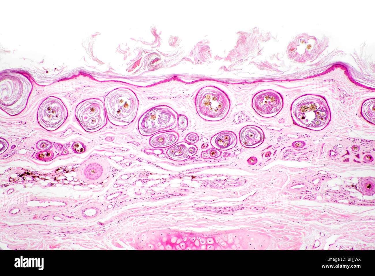
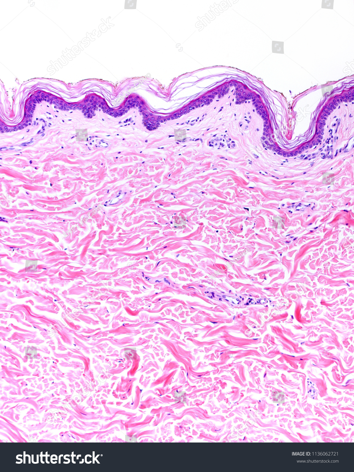
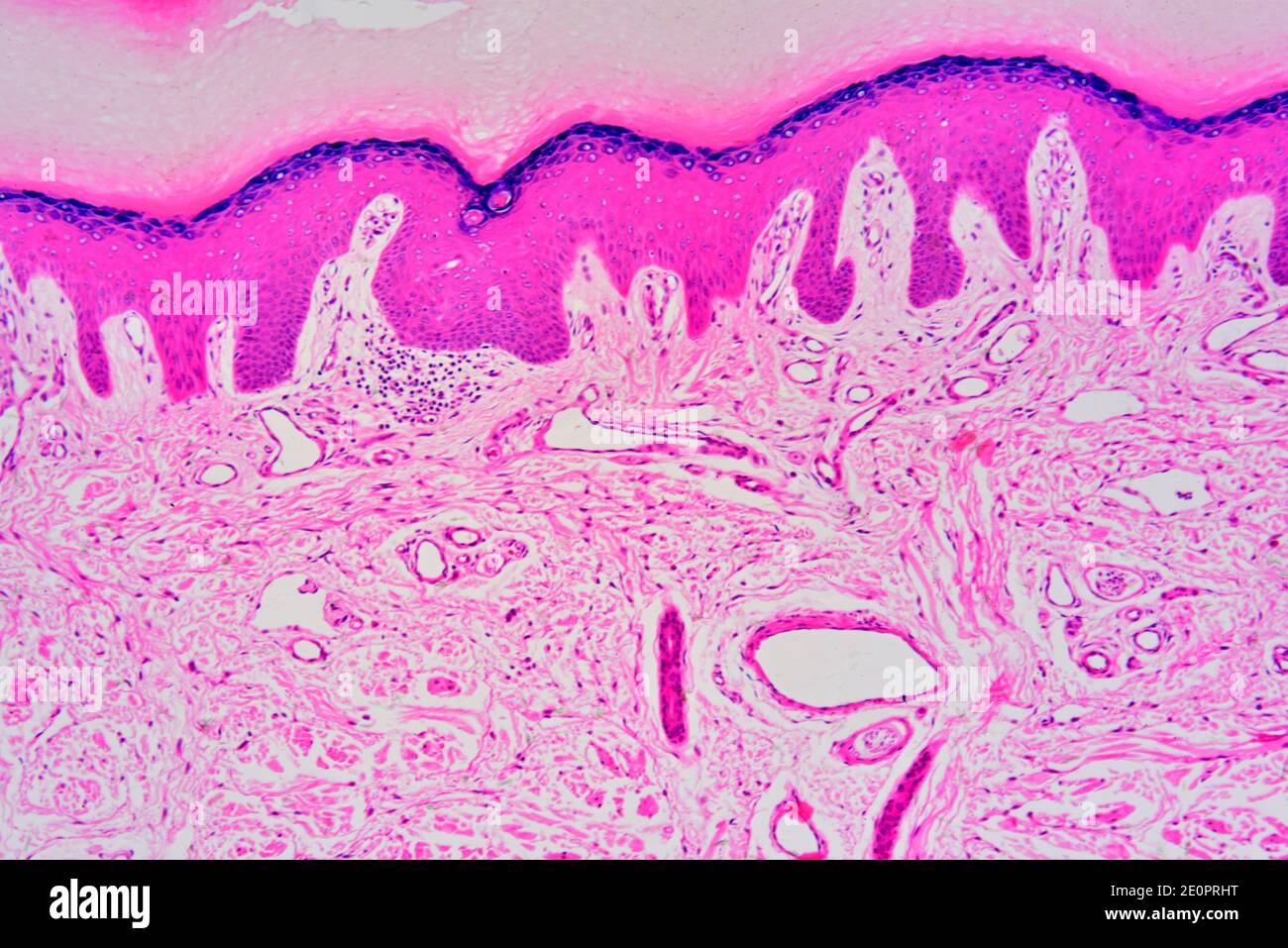
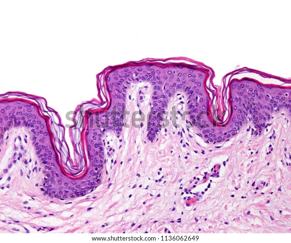
Thin Skin. Hematoxylin-eosin Stock Photo - Dreamstime Get 15 images free trial Thin skin. Hematoxylin-eosin Royalty-Free Stock Photo Epidermis of the thin skin. It can be identified the stratum basale, spinosum, a narrow stratum granulosum and a superficial well defined stratum corneum. The epidermis rest over the dermis stratum corneum, stratum granulosum, stratum basale, hematoxylin eosin, skin,

Photomicrograph of thin skin
Histology Skin Stock Photos, Pictures & Royalty-Free Images - iStock Thin skin Thin skin showing the epidermis with their different layers resting on dermis. Skin papilloma of a human Skin papilloma of a human, highly detailed segment of panorama. Photomicrograph as seen under the microscope, 10x zoom. Tissue types. connective, muscle, nervous, and epithelial cells Photomicrograph of Thick Skin Quiz - PurposeGames.com This is an online quiz called Photomicrograph of Thick Skin There is a printable worksheet available for download here so you can take the quiz with pen and paper. Your Skills & Rank Total Points 0 Get started! Today's Rank -- 0 Today 's Points One of us! Game Points 6 You need to get 100% to score the 6 points available Actions Add to Playlist Glossary of Stainless Steel Terms - Unified Alloys Skin A thin surface layer that is different from the main mass of a metal object, in composition, structure or other characteristics. Skull A layer of solidified metal or dross on the wall of a pouring vessel often when metal has been poured. Slab A piece of metal, intermediate between ingot and plate, at least twice as wide as it is thick.
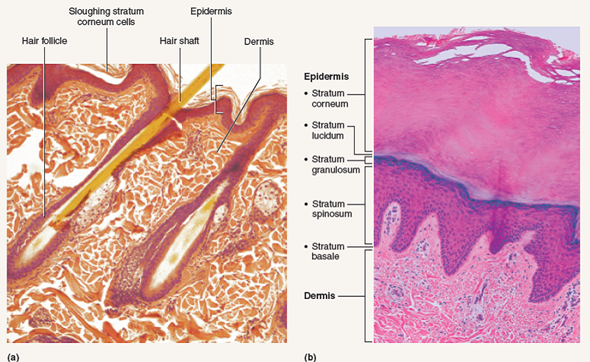
Photomicrograph of thin skin. Label The Photomicrograph Of Thick Skin. - Martina Eisenhower 1 answer to label the photomicrograph of thin skin. The epidermis, made of closely packed epithelial cells, and the dermis, made of dense, irregular connective tissue . Epidermis Of Thick Skin from eugraph.com The skin is composed of two main layers: Thick skin showing epithelial detail. Practice labeling the layers of the skin. unit 4 lab.docx - LAB Unit 4 EXERCISE 7: The Integumentary... FIGURE 7.5 Photomicrograph of the skin and accessory structures. • hair bulbs • hair follicle • hair root • papilla of hair • sebaceous gland 1 Sebaceous gland 2 Hair follicle 3 Hair root 4 Hair bulb 5 Papilla of hair SKIN | The Big Picture: Histology | AccessBiomedical Science | McGraw ... Epidermis in thick skin, the type of skin found on the palms, flexor surfaces of the digits, and the soles of feet, is 400 to 600-μm thick. In comparison, epidermis in thin skin, which is found everywhere else on the body, is only 75 to 150-μm thick. Figure 11-1: Layers of the Skin | Anatomy and Physiology I | | Course Hero The cells in all of the layers except the stratum basale are called keratinocytes. A keratinocyte is a cell that manufactures and stores the protein keratin. Keratin is an intracellular fibrous protein that gives hair, nails, and skin their hardness and water-resistant properties.The keratinocytes in the stratum corneum are dead and regularly slough away, being replaced by cells from the ...
PDF Quantitative microscopical and histochemical study of the skin of mice ... 2-A A computerized photomicrograph of longitudinal section in the non-exposed skin of mice, showing thin and sparse elastic fibers arranged in fibrillary pattern in the dermis. (Orcein. stain, X400) 2-C A computerized photomicrograph of longitudinal section in the non-exposed skin of mice, showing melanin Anatomy, Skin (Integument), Epidermis - StatPearls - NCBI Bookshelf Squamous cell carcinoma is cancer that arises from mutated keratinocytes, usually due to UV damage in individuals with Type I or II skin types (light skin, blue or green eyes, red or blonde hair, burn and never tan) and often appear as scaly, flaky, thick red patches that may bleed or even appear wart-like. This type of skin cancer can metastasize. PDF The Integumentary System - Holly H. Nash-Rule, PhD 2. The thickness of the skin can be attributed to the presence of a fifth epithelial layer, the stratum lucidum, and a thicker stratum corneum and dermis. Thick skin lacks hair follicles, arrector pili muscles, and seba-ceous glands that are present on thin skin of the scalp. Rebecca ferguson boobs | Andrea bang sexy ♥ Live Sex Cams & Chats Label the photomicrograph of thin skin. Martin x chris; Minecraft dragon pixel art; Nude female fit; Owl fairy lamp; Male slave tumblr; Eat creampie.story; Felix deon art; Kawaii anime christmas; Stravinsky nude
photomicrograph of thick skin Diagram | Quizlet photomicrograph of thick skin Diagram | Quizlet photomicrograph of thick skin + − Learn Test Match Created by mckennawebber Terms in this set (7) epidermis (stratum corneum - stratum basale) ... stratum corneum ... stratum lucidum ... stratum granulosum ... stratum spinosum ... stratum basale ... dermis ... Sets found in the same folder American Journal of Veterinary Research | AVMA This month's cover image is a photomicrograph of a skin sample obtained from the incision site of a koi 2 weeks after coelioscopy. The image depicts poor healing; the epithelium is incomplete, and the wound surface is partially covered by a layer of serofibrinous crust and cellular debris. Academia.edu - Cambridge International AS and A Level Biology ... • Metabolism: all the chemical processes occurring within an organism • Enzymes increase the rate of reactions that occur in living organisms. Question : Label the photomicrograph of thin skin. Dermis Duct of ... Expert Answer. 100% (37 ratings) A …. View the full answer. Transcribed image text: Label the photomicrograph of thin skin. Dermis Duct of sebaceous gland Hair Follicle Sebaceous gland Hair Epidermis.
View Image
Sensory nerve endings in the human female umbilical skin - LWW A photomicrograph of a section of the umbilical skin showing a relatively thin epidermis and a thin horny layer. Note the irregular short few dermal papillae. H&E ×100. ... A photomicrograph of the umbilical skin showing the Meissner's corpuscle (M) in a dermal papilla. It is tightly abutting the overlying basal layer of the epidermis.

Thin Skin Showing Epidermis Different Strata Stock Photo ...
Skin Histology Stock Photos, Pictures & Royalty-Free Images - iStock Thin skin Thin skin showing the epidermis with their different layers resting on dermis. Skin papilloma of a human Skin papilloma of a human, highly detailed segment of panorama. Photomicrograph as seen under the microscope, 10x zoom. Micrograph of squamous cell carcinoma of the head and neck

a) A photomicrograph of the section of thin skin tissue from ...
Mange in the Red Fox | Wildlife Online Summary: There are several different forms of mange, each caused by a different species of mite, but sarcoptic mange most commonly affects foxes. Sarcoptic mange is a skin disease caused by the small (2 to 4 mm, or less than one-quarter of an inch) parasitic mite Sarcoptes scabiei, several thousand of which may burrow into a single square-centimetre of skin.
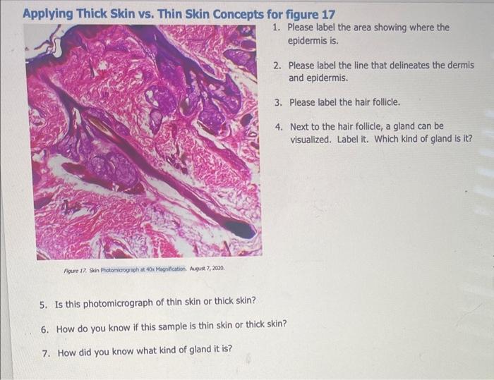
Applying Thick Skin vs. Thin Skin Concepts for figure | Chegg.com
Antimicrobial resistance - Wikipedia Clinical misuse by healthcare professionals is another cause leading to increased antimicrobial resistance. Studies done by the CDC show that the indication for treatment of antibiotics, choice of the agent used, and the duration of therapy was incorrect in up to 50% of the cases studied. In another study done in an intensive care unit in a major hospital in France, it was shown that …

Protection effects of rice protein hydrolysate on UVB ...
photomicrographs of thin skin Flashcards | Quizlet photomicrographs of thin skin. Term. 1 / 4. stratum corneum. Click the card to flip 👆. Definition. 1 / 4. ... Click the card to flip 👆.
![5 The Integumentary System. PSR #3 Cut- [skin] Derm ...](https://images.slideplayer.com/37/10718635/slides/slide_55.jpg)
5 The Integumentary System. PSR #3 Cut- [skin] Derm ...
Cambridge IGCSE Biology Third Edition Hodder Education Cambridge IGCSE Biology Third Edition Hodder Education

Topographic Relationships of the Peritoneal Canal of ...
Anatomy of the Epidermis with Pictures - Verywell Health The epidermis is composed of layers of skin cells called keratinocytes. Your skin has four layers of skin cells in the epidermis and an additional fifth layer in areas of thick skin. The four layers of cells, beginning at the bottom, are the stratum basale, stratum spinosum, stratum granulosum, and stratum corneum.
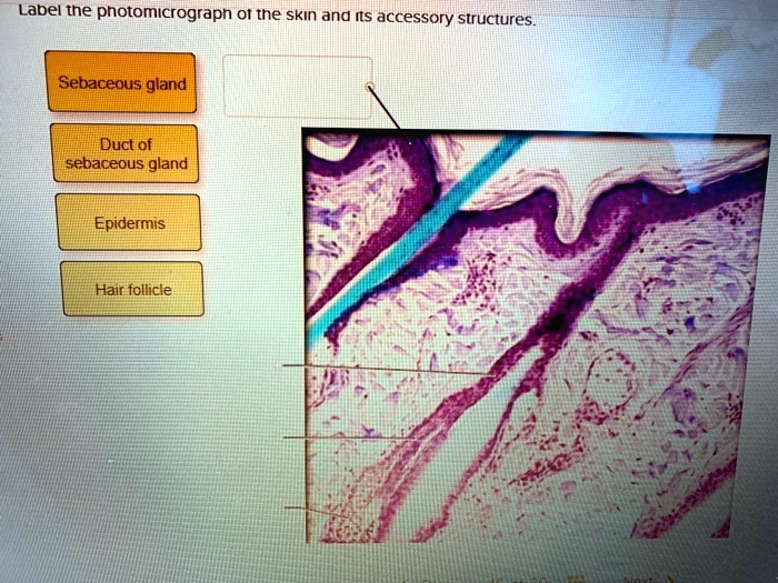
SOLVED: Label tne photomicrograph Of the Skin and Its ...
Cysts and cystic-appearing lesions of the knee: A pictorial essay A thin connection to the original tear can usually be demonstrated ... from direct extension of infection from adjacent tissues, like in osteomyelitis, or when the skin barrier has ... of the knee. (C) Coronal PDW image confirms that the mass (arrow) arises from common peroneal nerve (arrowheads). (D) Photomicrograph [hematoxylin ...
Skin cross section hi-res stock photography and images - Alamy
Leaf micrograph section hi-res stock photography and images - Alamy RF2JMKHPT - Epidermis of thin skin. From the depth to the surface it can be identified the the basale, spinosum, granulosum and corneum stra. RMG3YXYY - Brightfield photomicrograph, Sycamore leaf section TS showing central vein and cell structure.
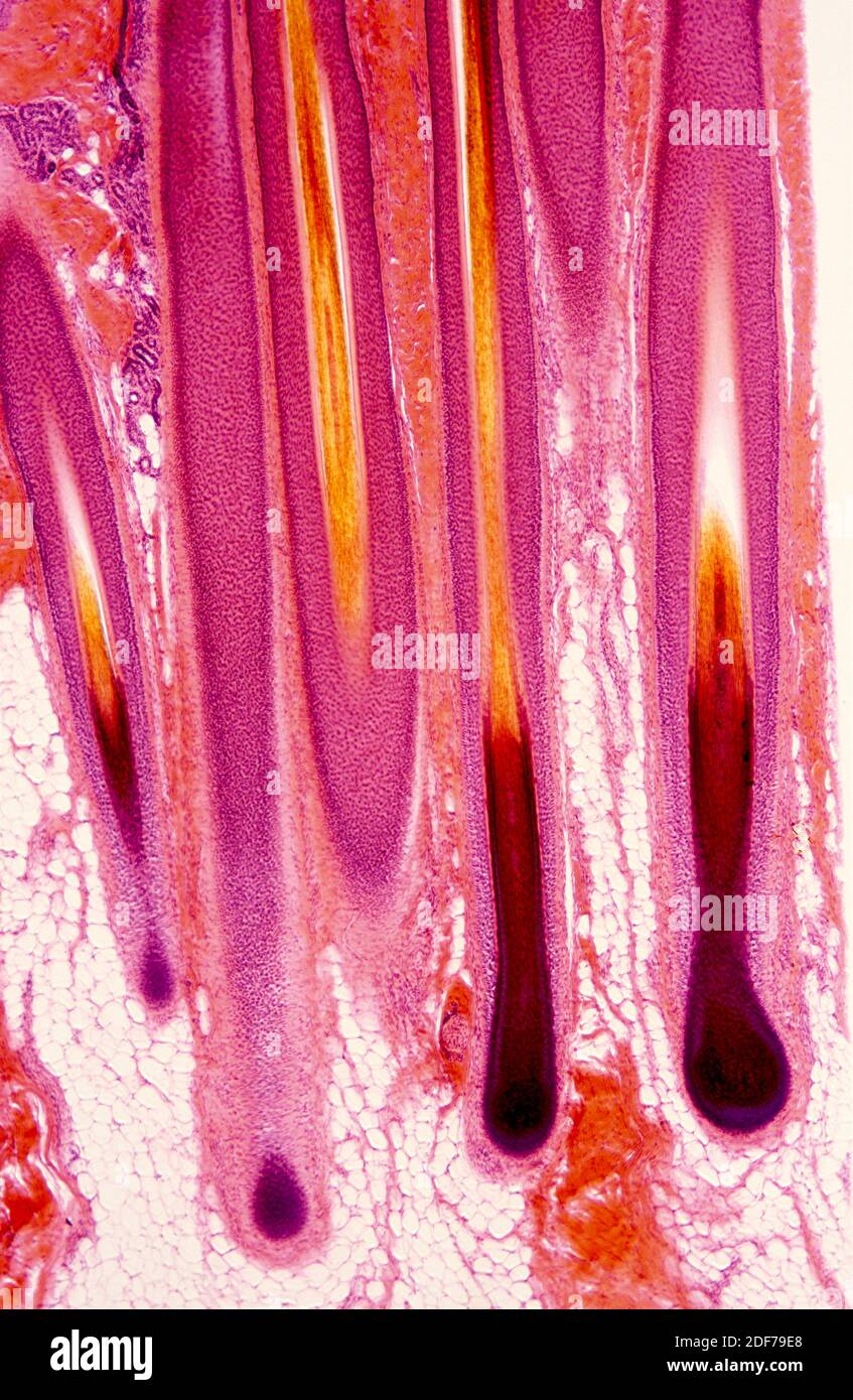
Human hair follicle hi-res stock photography and images - Alamy
Photomicrograph of Thin Skin - Printable - PurposeGames.com Photomicrograph of Thin Skin - Printable Download and print this quiz as a worksheet. You can modify it to fit your needs before you download. This is a printable worksheet made from a PurposeGames Quiz. To play the game online, visit Photomicrograph of Thin Skin Download Printable Worksheet Please note!

Presentation, Diagnostic Imaging, and Clinical Outcome of ...
Pilomatricoma as a Diagnostic Pitfall in Clinical Practice: Report of ... Pilomatricoma (PMC) is a relatively uncommon benign skin neoplasm arising from the skin adnexa. Since the first description of PMC in 1880, there has been a gradual increase in understanding of the morphologic features and clinical presentation of this tumor. ... Photomicrograph of pilomatricoma showing the characteristic basaloid (thick arrow ...

The skin of Diplometopon zarudnyi. Photomicrographs of ...
Label The Photomicrograph Of Thick Skin / Solved Label The ... - Blogger The epidermis of thick skin has five layers: Thick skin · stratum basale (also known as s. Label the photomicrograph of thick skin. It has a fifth layer,. Start studying photomicrograph of the epidermal layer in thick skin. The outer layer of cells in this micrograph is the thinnest layer and. A few layers of cells that are .

1,967 Skin Histology Stock Photos, Pictures & Royalty-Free ...
Cyanosite Image Gallery - Cyanobacteria Chloroflexus sp. - Wider view higher resolution image of specimen above. Cyanosarcina sp. from a pulp and paper waste-treatment system in Brazil. Cylindrospermopsis raciborskii - Heterocysts and akinetes visible. 400X. Very old laboratory culture of Cyanothece sp. forms solid roof to retain moisture.

Photomicrograph of Thick Skin Quiz
(PDF) The Developing Human-Clinically Oriented ... - Academia.edu Keith L. Moore

Collagen and elastic fibers. cajal-gallego trichrome. Dermis ...
Hair Under a Microscope - Rs' Science The fur is a thick growth of hair that covers the skin of many mammals. It consists of a combination of longer guard hair on top and shorter fleece hair (also known as underfur or down hair) beneath. The guard hair keeps moisture from reaching the skin; the underfur acts as an insulating blanket that keeps the animal warm. Thermal insulation
_Chr15/004_B10_cpdm_cont_8wk_fe_40X_index.jpg)
Skinbase - Mutant Mouse Skin Database
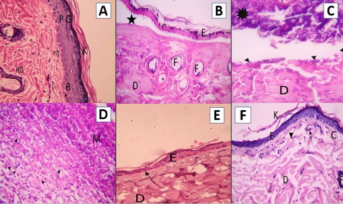
(a): A photomicrograph of the section of thin skin tissue from the ... (a): A photomicrograph of the section of thin skin tissue from the control group showing the epidermis layer and dermis differentiated into papillary (P) and reticular (R) layers. Note the hair...

View Image
Solved Label the photomicrograph of thick skin | Chegg.com Expert Answer. 91% (11 ratings) Transcribed image text: Label the photomicrograph of thick skin.
Longitudinal Comparison of Enzyme- and Laser-Treated ...
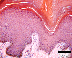
Basic histological structure and functions of facial skin Thin skin. Human. Light micrograph of a longitudinal section of female cheek showing the four layers of the thin skin and the four different cell types of epidermis. In panel A: Green degraded...

Thin Skin. Hematoxylin-eosin Stock Image - Image of ...
Bacillus anthracis - Wikipedia Bacillus anthracis is a gram-positive and rod-shaped bacterium that causes anthrax, a deadly disease to livestock and, occasionally, to humans.It is the only permanent pathogen within the genus Bacillus.Its infection is a type of zoonosis, as it is transmitted from animals to humans. It was discovered by a German physician Robert Koch in 1876, and became the first bacterium to …
Low Magnification Micrograph Human Thin Skin Stock Photo ...
Label The Photomicrograph Of Thick Skin - Faktor yang Label the photomicrograph of thick skin. 1 answer to label the photomicrograph of thin skin. The epidermis of thick skin has five layers: Hypodermis label the layers of the epidermis in thick skin in figure 7.2. A few layers of cells that are . Apocrine sweat gland label the photomicrograph in figure 7.4. Label the photomicrograph of thick skin.
Solved Figure 7.6: (a) Thin skin with hairs (120X). (b ...
Question : Question 31 points Label the photomicrograph of thin skin ... Question 31. A first grade teacher wishes to "shape" her student's writing of the alphabet. The teacher should: a, reward the child whenever the c... Question 31. A neurotransmitter that allows sodium ions to leak into a postsynaptic neuron causes: A) inhibitory postsynaptic damage to the myelin sheath C) excitatory postsynaptic...

OpenVetJ-10-431-g009.jpg
Glossary of Stainless Steel Terms - Unified Alloys Skin A thin surface layer that is different from the main mass of a metal object, in composition, structure or other characteristics. Skull A layer of solidified metal or dross on the wall of a pouring vessel often when metal has been poured. Slab A piece of metal, intermediate between ingot and plate, at least twice as wide as it is thick.

Dermis of thin skin. Hematoxylin-eosin
Photomicrograph of Thick Skin Quiz - PurposeGames.com This is an online quiz called Photomicrograph of Thick Skin There is a printable worksheet available for download here so you can take the quiz with pen and paper. Your Skills & Rank Total Points 0 Get started! Today's Rank -- 0 Today 's Points One of us! Game Points 6 You need to get 100% to score the 6 points available Actions Add to Playlist

Pin by nico x. on Anatomy | Games, Tetris, Anatomy
Histology Skin Stock Photos, Pictures & Royalty-Free Images - iStock Thin skin Thin skin showing the epidermis with their different layers resting on dermis. Skin papilloma of a human Skin papilloma of a human, highly detailed segment of panorama. Photomicrograph as seen under the microscope, 10x zoom. Tissue types. connective, muscle, nervous, and epithelial cells
American Journal of Case Reports | Lumbar Spinal Epidural ...
Solved Label these structures located in axillary skin. Hair ...

Collagen and Elastic Fibers. Cajal-Gallego Trichrome Stock ...
Skin: The Histology Guide

Chapter 5 The Integumentary System Shilla Chakrabarty, Ph.D ...
Epidermis human skin hi-res stock photography and images - Alamy
The Integumentary System | SpringerLink

Photomicrograph of a section in the skin of an albino rat ...

The erectile cheek-spine apparatus in the bristlenose catfish ...

Thin Skin. Hematoxylin-eosin Stock Photo - Image of ...
KoreaMed Synapse
Epidermis Thin Skin Can Be Identified Foto Stok 1136062649 ...
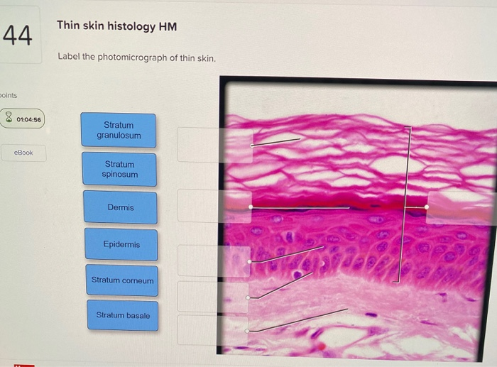
Solved Thin skin histology HM 44 Label the photomicrograph ...

A&P 1 Exercise_7 Activity 1 & 2 & RYK and UYK.docx - LAB ...

Pin by nico x. on Anatomy | Thick skin, Epidermis, Dermis
Histological assessment, anti-quorum sensing, and anti ...

Photomicrographs of skin (thin skin) Diagram | Quizlet
Chapter 5
Clearance of experimental cutaneous Staphylococcus aureus ...


Komentar
Posting Komentar